Showing posts with label waves. Show all posts

Sunday, February 10, 2019

Neural field models for latent state inference

The final paper from my Edinburgh postdoc in the Sanguinetti and Hennig labs (perhaps, we shall see). 

[get PDF]

We combined neural field modelling with point-process latent state inference. Neural field models capture collective population activity like oscillations and spatiotemporal waves. They make the simplifying assumption that neural activity can be summarized by the average firing rate in a region.

High-density electrode array recordings can now record developmental retinal waves in detail. We derived a neural field model for these waves from the microscopic model proposed by Hennig et al.. This model posits that retinal waves are supported by an quiescent, active, and refractory states. 

Fig 3. Spatial 3-state neural-field model exhibits self-organized multi-scale wave phenomena. Simulated example states at selected time-points on a [0,1]² unit interval using a 20×20 grid with effective population density of $\rho{=}50$ cells per unit area, and rate parameters $\sigma{=}0.075$, $\rho_a {=} 0.4$, $\rho_r {=} 3.2 \times 10^{−3}$, $\rho_e {=} 0.028$, and $\rho_q {=} 0.25$ (Methods: Sampling from the model). As, for instance, in neonatal retinal waves, spontaneous excitation of quiescent cells (blue) lead to propagating waves of activity (red), which establish localized patches in which cells are refractory (green) to subsequent wave propagation. Over time, this leads to diverse patterns of waves at a range of spatial scales. 

Tuesday, September 5, 2017

Inferring unobserved neural field intensities from spiking observations

Edit: I am very happy to report that this work has now been published in PLoS Computational Biology.

I'll be presenting our ongoing work on merging neural field models with statistical inference at the Integrated Systems Neuroscience Workshop in Manchester, and at the Bernstein Conference in Göttingen. [get poster PDF]

What's exciting about this work is that it combines modelling principles from statistical physics and statistical inference. We start with a detailed microscopic model, and then construct a second-order neural field model, which is then used directly for statistical inference. Normally, neural field models are only treated as abstract, qualitative mathematical models, and are rarely integrated with data. 


Video: Simulation of 3-state Quiescent-Active-Refractory blue, red, green) neural field model of spontaneous retinal waves that occur during development. Waves are generated by the inner retina, and drive retinal ganglion cell spiking, which we can observe on a high-density multi-electrode array. [get original avi from Github]

Saturday, February 4, 2017

System-size expansion and Gaussian moment closure for Quiescent-Active-Refractory model

Epilogue: These notes concern the system size expansion and moment closure later published as "Neural field models for latent state inference: Application to large-scale neuronal recordings[pdf] (more) 

These notes derive the Kramers-Moyal system size expansion and approximate equation for the evolution of means and covariances of a single-compartment neural mass model with Quiescent (Q), Active (A), and Refractory (R) states. The derivations here are identical to the standard ones for the Susceptible (S), Infected (I), and Recovered (R) (SIR) model commonly used in epidemiology. 

[get these notes as PDF]

Saturday, October 17, 2015

Diverse spatiotemporal dynamics in primate motor cortex local field potentials

Edit: this work has now been published as two papers. The first finds that mesoscopic beta-LFP oscillations may arise due to synchronization of rhythmic spiking in single neurons. The second  explores how changes in synchronization relate to the diverse patterns illustrated in the poster below.  

I'll present some of my ongoing thesis research at SfN as a poster this year. (This was originally titled "Identification of (~20 Hz) beta spatiotemporal dynamics in motor cortex LFPs".)

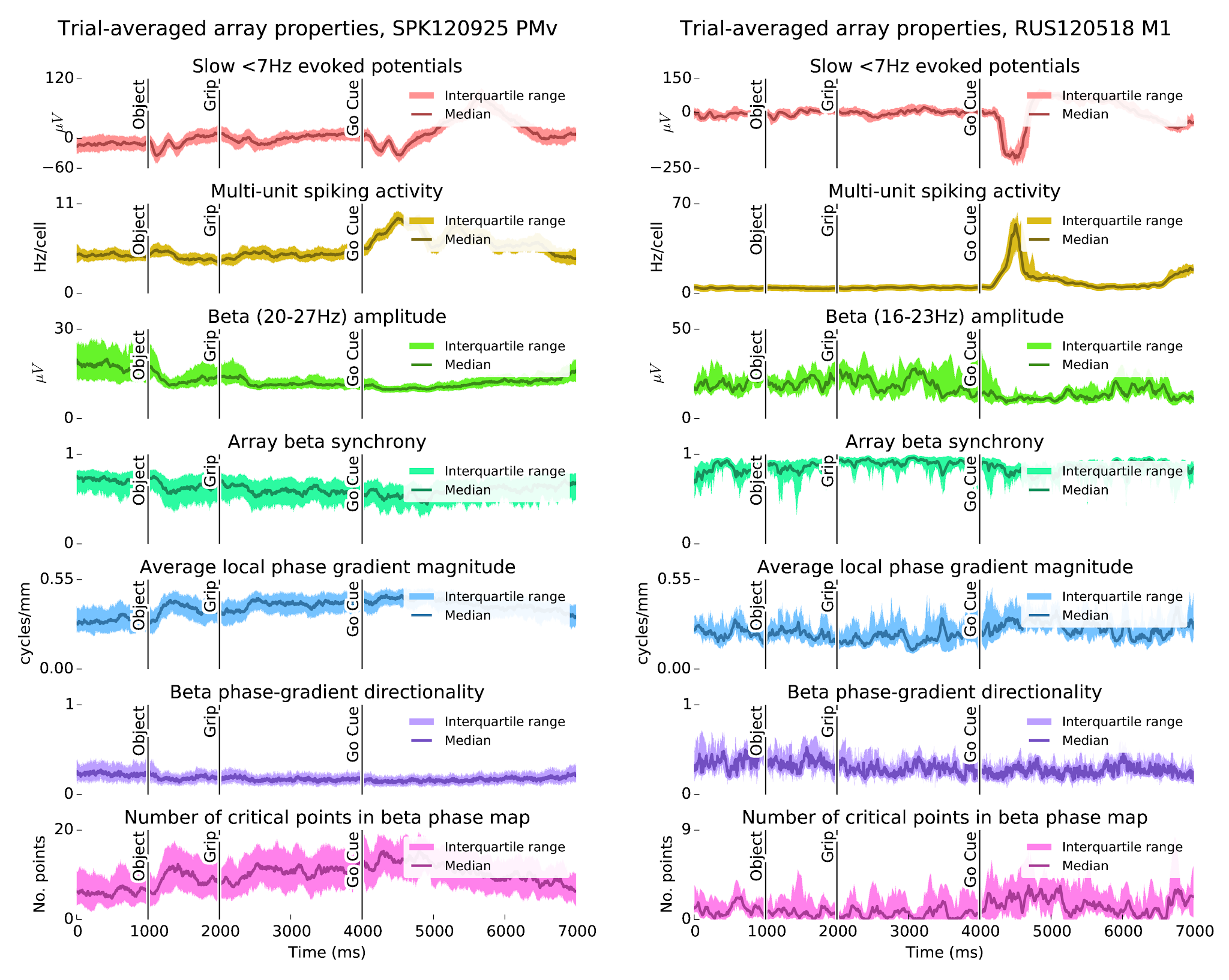
I've been looking into spatiotemporal waves in beta (~20 Hz) LFP oscillations during steady-state movement preparation. The wave dynamics seem to be more complex than previously reported [12]. Patterns vary depending on the properties of the beta-LFP oscillations, perhaps reflecting phase synchronization dynamics between local modules, or a signature of external input? 


Abstract:

Modulation of beta (10-45Hz) oscillations is a prominent feature of primate motor cortex. Beta power is typically elevated during movement preparation, suppressed around movement onset, and enhanced during isometric force tasks. As shown by previous studies, beta oscillations can also appears as traveling waves in primate motor cortex. Understanding the mechanisms underlying the rapid modulation of beta LFP activity and the associated spatiotemporal patterns may shed light on the functional roles of these oscillations. It may also have important implications for movement disorders where regulation of motor cortex beta activity is abnormal (e.g. Parkinson's disease). Here, we examine motor cortex beta spatiotemporal LFP activity using multielectrode arrays (MEAs) in m. mulatta during a cued reaching and grasping task with instructed delay. Data from two monkeys are analyzed, each with a 96-MEA in ventral premotor cortex (PMv), and two 48-MEAs in the primary motor cortex (M1) and dorsal premotor cortex (PMd), respectively. Our main findings are threefold: (1) The transient nature of beta oscillation events together with variations in the beta band center frequency makes the identification of spatiotemporal structures challenging. In particular, different filtering and preprocessing steps can alter the apparent spatiotemporal dynamics. (2) Furthermore, attempts to summarize wave dynamics in terms of simple global structures, like plane waves, or rotating (radiating) waves around (from) a critical point, may fail to meaningfully describe the full range of beta spatiotemporal activity. (3) Despite these challenges, we find a variety of beta spatiotemporal patterns ranging from asynchronous states, i.e. states with no clear wave dynamics, to more locally synchronized states with complex wave dynamics, to globally coherent states. These globally coherent states may exhibit either traveling wave dynamics or homogeneous synchrony. We conjecture that the transitions among these different patterns may result from fast modulations of the effective lateral connectivity or from changes in spatiotemporal inputs to the cortical area.

This poster can be cited as

Rule, M. E., Vargas-Irwin, C., Donoghue, J., Truccolo, W. (2015) Identification of (~20 Hz) beta spatiotemporal dynamics in motor cortex LFPs. [Poster] Society for Neuroscience 2015, Oct 19th, Chicago, Il, USA. 

Update: This work can now be found in the following papers

Rule, M.E., Vargas-Irwin, C.E., Donoghue, J.P. and Truccolo, W., 2017. Dissociation between sustained single-neuron spiking and transient β-LFP oscillations in primate motor cortexJournal of neurophysiology117(4), pp.1524-1543. 

Rule, M.E., Vargas-Irwin, C., Donoghue, J.P. and Truccolo, W., 2018. Phase reorganization leads to transient β-LFP spatial wave patterns in motor cortex during steady-state movement preparationJournal of neurophysiology119(6), pp.2212-2228.

Wednesday, April 1, 2015

Directional statistics for spatiotemporal wave analysis

I've been searching for a good distribution that can be used to summarize how the distribution of phases and amplitudes evolves during transient synchronization events of beta (~20 Hz) local field potentials (LFP) in motor cortex. So far, it seems difficult to find a single distribution family that works in all cases. 

Spatiotemporal wave activity in beta oscillations in motor cortex can be described in terms of the beta-band analytic LFP signal, which has both a magnitude and a phase, and who's real part is equal to the time-domain value of the beta-filtered signal. 

\begin{equation}z_k(t) = \beta_k (t) + i\cdot  \operatorname{Hilbert}(\beta_k)(t) = r_k(t) e^{i \theta_k(t)},\\\textrm{ where $k$ indexes over channels}\end{equation}

Circular statistics can be used to summarize the distribution of analytic signal phase, in order to detect synchrony and wave events.

[read more in the PDF


Figure 2: Neither the complex Gaussian nor log-polar statistics perfectly describe the distributions of analytic signal. In these plots, the black ellipse represents a complex Gaussian model of the data, with the ellipse boundary at one standard deviation, and the ellipse axes representing the eigenvectors of the covariance matrix $\Sigma$. The cyan contours represent a log-polar model of the data, which uses the mean and standard deviation of the log-amplitude, as well as the circular mean and standard deviation of the phases, to model the data in log-polar space. (a) When phase is concentrated, and not correlated with amplitude, both the log-polar statistics and the complex Gaussian distribution describe the data well. (b) When phase and amplitude are correlated, the log-polar model cannot capture the phase-amplitude dependence. (c) During traveling wave events, signal amplitude is high, and there is dispersion in phase. In these cases, the log-polar statistics are more appropriate than the complex Gaussian. (d) Traveling wave events appear to often evolve from states that show a mixture of synchrony and standing wave dynamics. The log-polar statistics break down when the phase distribution is bimodal, but the complex Gaussian can describe these states well. (e) At low signal amplitudes, the system is often asynchronous, and the phase and amplitude of the log-polar model are poorly defined. (f) Although rare or absent in our data, a hypothetical distribution with uniform phase and concentrated amplitude could occur, say, during traveling wave events with short wavelength. In this case, the complex Gaussian model is especially bad.

Friday, March 20, 2015

Three unbiased estimators of spike-field coherence

[get notes as PDF]

In these notes we show a simplified expression for the Pairwise Phase Consistency (PPC) measure of Vinck et al. (2010). We illustrate it's relation to a bias-corrected spike-field coherence measure from Grasse et al. (2010), and discuss an third notion of spike-field coherence that is intermediate between the two. 

Here, we use the term "event-triggered" rather than "spike-triggered", because we want to apply these measures to neural events other than spikes (e.g. beta-frequency transients in motor cortex).

Pairwise Phase Consistency

Vinck et al. (2010) define Pairwise Phase Consistency (PPC) as the expected dot product between all pairs of (spike-triggered) phase measurements. 

\[\hat\Upsilon = \frac 2 {N(N-1)} \sum_{j=1..N-1} \sum_{k=j+1..N} \cos(\theta_j - \theta_k)\]

There is an alternative way to express PPC that is faster to calculate, and also reveals a relationship between PPC and similar alternatives. 

Motor cortex LFP spatiotemporal dynamics in a cued grasp with instructed delay task

Update: Portions of these notes have now been published in the Journal of Neurophysiology as  "Dissociation between sustained single-neuron spiking and transient β-LFP oscillations in primate motor cortex" and "Phase reorganization leads to transient β-LFP spatial wave patterns in motor cortex during steady-state movement preparation".

[get notes as PDF]

Task-locked modulations in neural activity

The Cued Grasp with Instructed Delay (CGID) task reliably elicits task-locked activity in all three motor areas (M1, PMd, PMv).

  • Consistent with prior literature, the movement period of the CGID task is marked by slow motor evoked potentials (Fig. 2), increased single-unit firing rates (Fig. 3), and beta suppression (Fig. 4).
  • Beta oscillations are enhanced during the first four seconds of the task, although there are some differences between subjects.
  • The average level of beta-LFP synchrony is correlated with beta-LFP power, and varies across phases of the task.
  • We find no evidence of task-locked phase resetting of beta LFP oscillations
  • The spatiotemporal structure of beta-LFP waves is correlated with amplitude and synchrony, with lower amplitudes reflecting more complex wave structures, and higher amplitudes as more synchronous.

figure1

Figure 1: The CGID task reliably elicits evoked potentials, which correlate with beta suppression. In subject S, beta power is strongest in the first second before object presentation. In subject R, beta oscillations are more variable, with somewhat stronger power between the grip and go cues. In both animals, high beta power appears to correspond to periods of higher beta synchrony, and larger phase gradient directionality, a measure of how much LFP activity resembles a plane wave. Conversely, increases in the average magnitude of the Hilbert phase gradient, which summarizes how quickly beta phase changes over the array, and in the number of critical points in the Hilbert phase gradient, which summarizes the complexity of the beta spatiotemporal wave patterns, correspond to periods of beta suppression.