Showing posts with label m1. Show all posts

Thursday, June 14, 2018

Phase reorganization leads to transient β-LFP spatial wave patterns in motor cortex

The final paper from my thesis is, at long last, published [get PDF]. 

We studied traveling waves observed in electrical Local Field Potential (LFP) signals in primate motor cortex. We found that the structure of traveling waves in beta LFP oscillations was more complex than previously appreciated. 

Previous theoretical work noted that traveling waves in the brain do not always reflect "true" traveling waves, like the ripples from throwing a stone into water: They can also arise from common inputs arriving at different times, or from transient spatial reorganization of coupled oscillators.

We sought to clarify which of these scenarios is consistent with beta-LFP traveling waves in motor cortex. Our previous work looking at the role of single neurons suggested that the waves may be phase waves in coupled oscillators, rather than traveling pulses from a defined source.

This study supports the phase wave scenario. We also discovered a rich variety of spatial structures not previously reported, such as spiral and radiating waves and more complex structures. 

Many thanks to Carlos Vargas-Irwin, John P. Donoghue, and Wilson Truccolo. This work can be cited as

Rule, M.E., Vargas-Irwin, C., Donoghue, J.P. and Truccolo, W., 2018. Phase reorganization leads to transient β-LFP spatial wave patterns in motor cortex during steady-state movement preparation. Journal of neurophysiology, 119(6), pp.2212-2228. 

Figure 3, surveying the variety of patterns:

Fig. 3. Transient beta-local field potential (β-LFP) oscillations exhibit a rich variety of spatiotemporal wave patterns. In addition to traveling plane waves, beta spatiotemporal dynamics showed synchronous states, radiating and rotating waves, and other more complex wave patterns. Each example was taken from the 4×4-mm² area sampled by the 10×10 multielectrode array in ventral premotor cortex area of subject S. Missing electrodes were interpolated from nearest neighbors. Average phase-delay maps were computed by wrapping Hilbert phases at the median frequency of the wave event before computing the average analytic signal. The mean analytic signal was smoothed at a 2-mm scale to generate the phase-delay maps pictured. The smoothed Hilbert phase (φ) was differentiated to extract critical points from the wave dynamics, shown as a blue dot for a radiating wave and red dots for rotating waves. Spatially synchronized states were detected as patterns where the angular distribution of analytic signals was concentrated as shown by the first (top to bottom) example. Plane-wave states were detected as spatial patterns where the angular distribution of the phase gradient (∇φ) direction was concentrated, as shown by the second example.

Thursday, January 26, 2017

Dissociation between sustained single-neuron spiking and transient β-LFP oscillations in primate motor cortex

Chapter two of my thesis has just been published! Rule et al. 2017 [PDF] explores the neurophysiology of beta (β) oscillations in primates, especially how single-neuron activity relates to population activity reflected in local field potentials (a.k.a. "brain waves").

Beta (~20 Hz) oscillations occur in frontal cortex. We've known about them for about a century, but still don't understand how they work or what they do. β-wave activity is related to "holding steady", so to speak. 

Beta oscillations are dysregulated in Parkinson's, in which movements are slowed or stopped. Beta oscillations are also reduced relative to slow-wave activity in ADHD, a disorder associated with motor restlessness and hyperactivity.

I looked at beta oscillations during movement preparation, where they seem to play a role in stabilizing a planned movement. I found that single neurons had very little relationship to the β-LFP brain waves. However! This appears to be for a good reason: the firing frequencies of neurons store information about the upcoming movement, and neurons firing at different frequencies cannot phase-lock together into a coherent population oscillation.

Anyone who's played in an orchestra knows that when notes are just slightly out of tune, you get interference patterns called beats. The same thing is happening in the brain, where many neurons firing at slightly different "pitches" cause β-LFP fluctuations, even though the underlying neural activity is constant.

This result provides a new explanation for how β-waves can appear as "transients" during motor steady-state: the fluctuations are cased by "beating", rather than changes in the β activity in the individual neurons. This differs from the prevailing theory for the origin of β transients in more posterior brain regions.

Many thanks to Carlos Vargas-Irwin, John Donoghue, and Wilson Truccolo. You can grab the PDF here. Please cite as

Rule, M.E., Vargas-Irwin, C.E., Donoghue, J.P. and Truccolo, W., 2017. Dissociation between sustained single-neuron spiking and transient β-LFP oscillations in primate motor cortex. Journal of neurophysiology, 117(4), pp.1524-1543.


Monday, June 6, 2016

Collective neural dynamics in primate motor cortex

As of the 29th of May, 2016, I officially have a Ph.D. in neuroscience! The thesis, Collective Neural Dynamics in Primate Motor Cortex, is available from the Brown University library [PDF].

I studied how single-neuron activity relates to large-scale collective neural dynamics during movement planning and execution. The thesis covers three research projects, which have been (will be) published as stand-alone papers:

  • Chapter 2, pp. 88-121: Contribution of LFP dynamics to spiking variability in motor cortex during movement execution. read more...
Rule, M.E., Vargas-Irwin, C., Donoghue, J.P. and Truccolo, W., 2015. Contribution of LFP dynamics to single-neuron spiking variability in motor c ortex during movement execution. Frontiers in systems neuroscience, 9, p.89.
  • Chapter 3, pp. 122:168: Dissociation between single-neuron spiking β-rhythmicity and transient β-LFP oscillations during movement preparation in primate motor cortex. read more…
Rule, M.E., Vargas-Irwin, C.E., Donoghue, J.P. and Truccolo, W., 2017. Dissociation between sustained single-neuron spiking and transient β-LFP oscillations in primate motor cortex. Journal of neurophysiology, 117(4), pp.1524-1543.
  • Chapter 4, pp. 169-213: Phase diversity and spatiotemporal wavedynamics in primate motor cortex local field potentials. read more…
Rule, M.E., Vargas-Irwin, C., Donoghue, J.P. and Truccolo, W., 2018. Phase reorganization leads to transient β-LFP spatial wave patterns in motor cortex during steady-state movement preparation. Journal of neurophysiology, 119(6), pp.2212-2228.

The introduction contains background on primate motor cortex (Chapter 1, pp. 7-88), including its constituent areas, how they connect with the rest of the brain, and how neurons connect to each-other within each area. It surveys what is known (as of 2016) about motor cortex population dynamics, LFP oscillations, and spatiotemporal waves. The section on statistical methods (Chapter 1.5, pp. 61-87) provides background for signal processing to extract single-neuron spikes and LFPs from multi-electrode array recordings. It also covers how to apply Generalized Linear Point-Process Models (PP-GLM) to analyze spiking neural data.

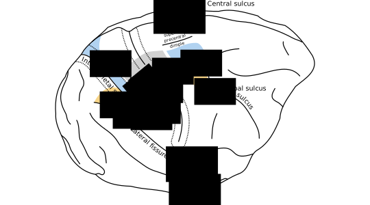
I'd also like to share two new illustrations from the introduction not published elsewhere:

Figure 1.1 

(high resolution PDF, SVG)


Figure 1.1: Anatomy of visually-guided reaching and grasping. During visually guided reaching and grasping, the arm and hand area of M1 coordinates with the dorsal and ventral premotor areas PMd and PMv. In this illustration, reciprocally connected motor and parietal areas are shaded in common colors. Premotor areas receive segregated streams of visual information from parietal cortex. Area PMd receives information about spatial geometry important for reaching from dorsal parietal areas (shaded in blue). Area PMv receives information about object geometry important for grasping from the parietal areas shaded in orange. Area M1 also receives feedback from somatosensory cortex (areas 3a,1,2, shaded in grey). Connections between parietal and premotor cortex are taken from Tanné-Gariépy et al. (2002), and anatomical boundaries of premotor areas are taken from Dum and Strick (2002).

Saturday, October 17, 2015

Diverse spatiotemporal dynamics in primate motor cortex local field potentials

Edit: this work has now been published as two papers. The first finds that mesoscopic beta-LFP oscillations may arise due to synchronization of rhythmic spiking in single neurons. The second  explores how changes in synchronization relate to the diverse patterns illustrated in the poster below.  

I'll present some of my ongoing thesis research at SfN as a poster this year. (This was originally titled "Identification of (~20 Hz) beta spatiotemporal dynamics in motor cortex LFPs".)

I've been looking into spatiotemporal waves in beta (~20 Hz) LFP oscillations during steady-state movement preparation. The wave dynamics seem to be more complex than previously reported [1, 2]. Patterns vary depending on the properties of the beta-LFP oscillations, perhaps reflecting phase synchronization dynamics between local modules, or a signature of external input? 


Abstract:

Modulation of beta (10-45Hz) oscillations is a prominent feature of primate motor cortex. Beta power is typically elevated during movement preparation, suppressed around movement onset, and enhanced during isometric force tasks. As shown by previous studies, beta oscillations can also appears as traveling waves in primate motor cortex. Understanding the mechanisms underlying the rapid modulation of beta LFP activity and the associated spatiotemporal patterns may shed light on the functional roles of these oscillations. It may also have important implications for movement disorders where regulation of motor cortex beta activity is abnormal (e.g. Parkinson's disease). Here, we examine motor cortex beta spatiotemporal LFP activity using multielectrode arrays (MEAs) in m. mulatta during a cued reaching and grasping task with instructed delay. Data from two monkeys are analyzed, each with a 96-MEA in ventral premotor cortex (PMv), and two 48-MEAs in the primary motor cortex (M1) and dorsal premotor cortex (PMd), respectively. Our main findings are threefold: (1) The transient nature of beta oscillation events together with variations in the beta band center frequency makes the identification of spatiotemporal structures challenging. In particular, different filtering and preprocessing steps can alter the apparent spatiotemporal dynamics. (2) Furthermore, attempts to summarize wave dynamics in terms of simple global structures, like plane waves, or rotating (radiating) waves around (from) a critical point, may fail to meaningfully describe the full range of beta spatiotemporal activity. (3) Despite these challenges, we find a variety of beta spatiotemporal patterns ranging from asynchronous states, i.e. states with no clear wave dynamics, to more locally synchronized states with complex wave dynamics, to globally coherent states. These globally coherent states may exhibit either traveling wave dynamics or homogeneous synchrony. We conjecture that the transitions among these different patterns may result from fast modulations of the effective lateral connectivity or from changes in spatiotemporal inputs to the cortical area.

This poster can be cited as

Rule, M. E., Vargas-Irwin, C., Donoghue, J., Truccolo, W. (2015) Identification of (~20 Hz) beta spatiotemporal dynamics in motor cortex LFPs. [Poster] Society for Neuroscience 2015, Oct 19th, Chicago, Il, USA. 

Update: This work can now be found in the following papers

Rule, M.E., Vargas-Irwin, C.E., Donoghue, J.P. and Truccolo, W., 2017. Dissociation between sustained single-neuron spiking and transient β-LFP oscillations in primate motor cortex. Journal of neurophysiology, 117(4), pp.1524-1543. 

Rule, M.E., Vargas-Irwin, C., Donoghue, J.P. and Truccolo, W., 2018. Phase reorganization leads to transient β-LFP spatial wave patterns in motor cortex during steady-state movement preparation. Journal of neurophysiology, 119(6), pp.2212-2228.

Wednesday, July 8, 2015

Contribution of LFP dynamics to single-neuron spiking variability in motor cortex during movement execution

My first Ph.D. publication is out! Contribution of LFP dynamics to single-neuron spiking variability in motor cortex during movement execution explores how the activity of individual neurons in motor cortex is related to population activity, as measured by electrical Local Field Potentials (LFPs). 

[get PDF] 

How do the actions of individual cells combine to create the emergent dynamics that underlie perception, thought, and action? To answer this question, we should study populations of single neurons, and ask how their activity is related to measures of collective population dynamics. This study was a collaboration between the Truccolo and Donoghue labs, and looked at neural population recordings from primate motor cortex during movement.

We found that the activity of single cells was tightly coupled to population activity as measured by LFPs, and that both of these signals were closely realated to movement. This suggest that, during movement execution, collective dynamics reflected in motor cortex LFPs mostly reflect the sensorimotor processes directly controlling movement output. It also suggests that primary motor cortex isn't engaged in other activities like cognition or future planning, while executing movements.

Importantly, we considered both past and future movement in this analysis, and found that single neurons and LFPs both contain information about recent and upcoming movements. This is consistent with the view that motor cortex acts as a dynamical pattern generator.

Many thanks to Carlos Vargas-Irwin, John P. Donoghue, and Wilson Truccolo. The article is open access, and you can also grab the PDF from Github. The paper can be cited as:

Rule, M.E., Vargas-Irwin, C., Donoghue, J.P. and Truccolo, W., 2015. Contribution of LFP dynamics to single-neuron spiking variability in motor cortex during movement execution. Frontiers in systems neuroscience, 9, p.89. 


Figure 4. Breakdown of LFP predictive power by frequency band and LFP feature. Box-plots over the population of isolated units (all sessions combined) showing the predictive power of models based on phase, amplitude, or analytic signal features in isolation from each of eight LFP bands. To better assess the individual predictive power of each LFP feature, models were fitted for each feature separately. Certain features, such as the instantaneous phase and analytic signal for the 0.3–2 Hz band, as well as the analytic signal amplitude modulation above 100 Hz, consistently predict spiking across all animals and areas.