Showing posts with label science. Show all posts

Friday, November 1, 2019

Causes and consequences of representational drift

Drs. Harvey, O'Leary, and myself, have just published a review in Current Opinion in Neurobiology titled the "Causes and consequences of representational drift" [PDF].

We explore recent results from the work of Alon Rubin and Liron Sheintuch in the Ziv lab and Laura Driscoll in the Harvey lab. Their work has shown that neural representations reconfigure even for fixed, learned tasks. 

We discuss ways that the brain might support this reconfiguration without forgetting what it has already learned. There might be a subset of stable neurons that maintain memories. Alternatively, neural representations can be highly redundant, and support a stable readout even as single cells seem to change. 

Finally, we conjecture that redundancy allows different brain areas to "error-correct" each other, allowing the brain to keep track of the shifting meaning of single neurons in plastic representations.

For a brief preview, here are the pre-publication versions of figures 2 and 3:

Figure 2:

Figure 2: Internal representations have unconstrained degrees of freedom that allow drift. (a) Nonlinear dimensionality reduction of population activity recovers the low-dimensional structure of the T-maze in Driscoll et al. (2017). Each point represents a single time-point of population activity, and is colored according to location in the maze. (b) Point clouds illustrate low-dimensional projections of neural activity as in (a). Although unsupervised dimensionality-reduction methods can recover the task structure on each day, the way in which this structure is encoded in the population can change over days to weeks. (c) Left: Neural populations can encode information in relative firing rates and correlations, illustrated here as a sensory variable encoded in the sum of two neural signals ($y_1 + y_2$). Points represent neural activity during a repeated presentation of the same stimulus. Variability orthogonal to this coding axis does not disrupt coding, but could appear as drift in experiments if it occurred on slow timescales. Right: Such distributed codes may be hard to read-out from recorded subpopulations (e.g. $y_1$ or $y_2$ alone; black), especially if they entail correlations between brain areas. (d) Left: External covariates may exhibit context-dependent relationships. Each point here reflects a neural population state at a given time-point. The relationship between directions $x_1$ and $x_2$ changes depending on context (cyan versus red). Middle: Internally, this can be represented a mixture model, in which different subspaces are allocated to encode each context, and the representations are linearly-separable (gray plane). Right: The expanded representation contains two orthogonal subspaces that each encode a separate, context- dependent relationship. This dimensionality expansions increases the degrees of freedom in internal representations, thereby increasing opportunities for drift. 

Thursday, June 14, 2018

Phase reorganization leads to transient β-LFP spatial wave patterns in motor cortex

The final paper from my thesis is, at long last, published [get PDF]. 

We studied traveling waves observed in electrical Local Field Potential (LFP) signals in primate motor cortex. We found that the structure of traveling waves in beta LFP oscillations was more complex than previously appreciated. 

Previous theoretical work noted that traveling waves in the brain do not always reflect "true" traveling waves, like the ripples from throwing a stone into water: They can also arise from common inputs arriving at different times, or from transient spatial reorganization of coupled oscillators.

We sought to clarify which of these scenarios is consistent with beta-LFP traveling waves in motor cortex. Our previous work looking at the role of single neurons suggested that the waves may be phase waves in coupled oscillators, rather than traveling pulses from a defined source.

This study supports the phase wave scenario. We also discovered a rich variety of spatial structures not previously reported, such as spiral and radiating waves and more complex structures. 

Many thanks to Carlos Vargas-Irwin, John P. Donoghue, and Wilson Truccolo. This work can be cited as

Rule, M.E., Vargas-Irwin, C., Donoghue, J.P. and Truccolo, W., 2018. Phase reorganization leads to transient β-LFP spatial wave patterns in motor cortex during steady-state movement preparation. Journal of neurophysiology, 119(6), pp.2212-2228. 

Figure 3, surveying the variety of patterns:

Fig. 3. Transient beta-local field potential (β-LFP) oscillations exhibit a rich variety of spatiotemporal wave patterns. In addition to traveling plane waves, beta spatiotemporal dynamics showed synchronous states, radiating and rotating waves, and other more complex wave patterns. Each example was taken from the 4×4-mm² area sampled by the 10×10 multielectrode array in ventral premotor cortex area of subject S. Missing electrodes were interpolated from nearest neighbors. Average phase-delay maps were computed by wrapping Hilbert phases at the median frequency of the wave event before computing the average analytic signal. The mean analytic signal was smoothed at a 2-mm scale to generate the phase-delay maps pictured. The smoothed Hilbert phase (φ) was differentiated to extract critical points from the wave dynamics, shown as a blue dot for a radiating wave and red dots for rotating waves. Spatially synchronized states were detected as patterns where the angular distribution of analytic signals was concentrated as shown by the first (top to bottom) example. Plane-wave states were detected as spatial patterns where the angular distribution of the phase gradient (∇φ) direction was concentrated, as shown by the second example.

Monday, January 1, 2018

Autoregressive point-processes as latent state-space models

In 2016 I started a postdoc with the labs of Guido Sanguinetti and Matthias H. Hennig—and this is the first paper to result!

[get PDF]

A central challenge in neuroscience is understanding how the activity of single cells combines to create the collective dynamics that underlie perception, cognition, and behavior. One way to study this is to build detailed models, and "coarse grain" them to see which details are important. 

Our paper develops ways to relate detailed point-process models to coarse-grained quantities, like average neuronal firing rates and correlations. Point-process models are used for statistical modelling of spike train data. They can reveal effective neural dynamics by capturing how neurons in a population inhibit or excite each-other and themselves. 

Preview of figures: 

Figure 2: Moment closure of autoregressive PPGLMs combines aspects of three modeling approaches:

(A) Log-linear autoregressive PPGLM framework (e.g., Weber & Pillow, 2017). Dependence on the history of both extrinsic covariates x(t) and the process itself y(t) are mediated by linear filters, which are combined to predict the instantaneous log intensity of the process. (B) Latent state-space models learn a hidden dynamical system, which can be driven by both extrinsic covariates and spiking outputs. Such models are often fit using expectation- maximization, and the learned dynamics are descriptive. (C) Moment closure recasts autoregressive PPGLMs as state-space models. History dependence of the process is subsumed into the state-space dynamics, but the latent states retain a physical interpretation as moments of the process history (dashed arrow). (D) Compare to neural mass and neural field models, which define dynamics on a state space with a physical interpretation as moments of neural population activity

Thursday, January 26, 2017

Dissociation between sustained single-neuron spiking and transient β-LFP oscillations in primate motor cortex

Chapter two of my thesis has just been published! Rule et al. 2017 [PDF] explores the neurophysiology of beta (β) oscillations in primates, especially how single-neuron activity relates to population activity reflected in local field potentials (a.k.a. "brain waves").

Beta (~20 Hz) oscillations occur in frontal cortex. We've known about them for about a century, but still don't understand how they work or what they do. β-wave activity is related to "holding steady", so to speak. 

Beta oscillations are dysregulated in Parkinson's, in which movements are slowed or stopped. Beta oscillations are also reduced relative to slow-wave activity in ADHD, a disorder associated with motor restlessness and hyperactivity.

I looked at beta oscillations during movement preparation, where they seem to play a role in stabilizing a planned movement. I found that single neurons had very little relationship to the β-LFP brain waves. However! This appears to be for a good reason: the firing frequencies of neurons store information about the upcoming movement, and neurons firing at different frequencies cannot phase-lock together into a coherent population oscillation.

Anyone who's played in an orchestra knows that when notes are just slightly out of tune, you get interference patterns called beats. The same thing is happening in the brain, where many neurons firing at slightly different "pitches" cause β-LFP fluctuations, even though the underlying neural activity is constant.

This result provides a new explanation for how β-waves can appear as "transients" during motor steady-state: the fluctuations are cased by "beating", rather than changes in the β activity in the individual neurons. This differs from the prevailing theory for the origin of β transients in more posterior brain regions.

Many thanks to Carlos Vargas-Irwin, John Donoghue, and Wilson Truccolo. You can grab the PDF here. Please cite as

Rule, M.E., Vargas-Irwin, C.E., Donoghue, J.P. and Truccolo, W., 2017. Dissociation between sustained single-neuron spiking and transient β-LFP oscillations in primate motor cortex. Journal of neurophysiology, 117(4), pp.1524-1543.


Monday, June 6, 2016

Collective neural dynamics in primate motor cortex

As of the 29th of May, 2016, I officially have a Ph.D. in neuroscience! The thesis, Collective Neural Dynamics in Primate Motor Cortex, is available from the Brown University library [PDF].

I studied how single-neuron activity relates to large-scale collective neural dynamics during movement planning and execution. The thesis covers three research projects, which have been (will be) published as stand-alone papers:

  • Chapter 2, pp. 88-121: Contribution of LFP dynamics to spiking variability in motor cortex during movement execution. read more...
Rule, M.E., Vargas-Irwin, C., Donoghue, J.P. and Truccolo, W., 2015. Contribution of LFP dynamics to single-neuron spiking variability in motor c ortex during movement execution. Frontiers in systems neuroscience, 9, p.89.
  • Chapter 3, pp. 122:168: Dissociation between single-neuron spiking β-rhythmicity and transient β-LFP oscillations during movement preparation in primate motor cortex. read more…
Rule, M.E., Vargas-Irwin, C.E., Donoghue, J.P. and Truccolo, W., 2017. Dissociation between sustained single-neuron spiking and transient β-LFP oscillations in primate motor cortex. Journal of neurophysiology, 117(4), pp.1524-1543.
  • Chapter 4, pp. 169-213: Phase diversity and spatiotemporal wavedynamics in primate motor cortex local field potentials. read more…
Rule, M.E., Vargas-Irwin, C., Donoghue, J.P. and Truccolo, W., 2018. Phase reorganization leads to transient β-LFP spatial wave patterns in motor cortex during steady-state movement preparation. Journal of neurophysiology, 119(6), pp.2212-2228.

The introduction contains background on primate motor cortex (Chapter 1, pp. 7-88), including its constituent areas, how they connect with the rest of the brain, and how neurons connect to each-other within each area. It surveys what is known (as of 2016) about motor cortex population dynamics, LFP oscillations, and spatiotemporal waves. The section on statistical methods (Chapter 1.5, pp. 61-87) provides background for signal processing to extract single-neuron spikes and LFPs from multi-electrode array recordings. It also covers how to apply Generalized Linear Point-Process Models (PP-GLM) to analyze spiking neural data.

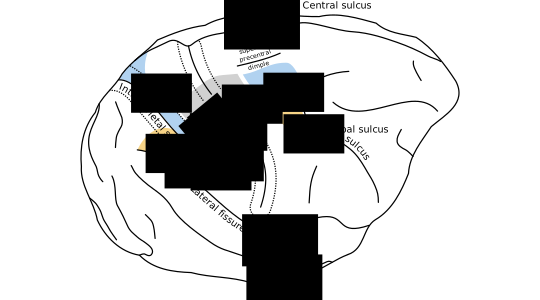
I'd also like to share two new illustrations from the introduction not published elsewhere:

Figure 1.1 

(high resolution PDF, SVG)


Figure 1.1: Anatomy of visually-guided reaching and grasping. During visually guided reaching and grasping, the arm and hand area of M1 coordinates with the dorsal and ventral premotor areas PMd and PMv. In this illustration, reciprocally connected motor and parietal areas are shaded in common colors. Premotor areas receive segregated streams of visual information from parietal cortex. Area PMd receives information about spatial geometry important for reaching from dorsal parietal areas (shaded in blue). Area PMv receives information about object geometry important for grasping from the parietal areas shaded in orange. Area M1 also receives feedback from somatosensory cortex (areas 3a,1,2, shaded in grey). Connections between parietal and premotor cortex are taken from Tanné-Gariépy et al. (2002), and anatomical boundaries of premotor areas are taken from Dum and Strick (2002).

Wednesday, July 8, 2015

Contribution of LFP dynamics to single-neuron spiking variability in motor cortex during movement execution

My first Ph.D. publication is out! Contribution of LFP dynamics to single-neuron spiking variability in motor cortex during movement execution explores how the activity of individual neurons in motor cortex is related to population activity, as measured by electrical Local Field Potentials (LFPs). 

[get PDF] 

How do the actions of individual cells combine to create the emergent dynamics that underlie perception, thought, and action? To answer this question, we should study populations of single neurons, and ask how their activity is related to measures of collective population dynamics. This study was a collaboration between the Truccolo and Donoghue labs, and looked at neural population recordings from primate motor cortex during movement.

We found that the activity of single cells was tightly coupled to population activity as measured by LFPs, and that both of these signals were closely realated to movement. This suggest that, during movement execution, collective dynamics reflected in motor cortex LFPs mostly reflect the sensorimotor processes directly controlling movement output. It also suggests that primary motor cortex isn't engaged in other activities like cognition or future planning, while executing movements.

Importantly, we considered both past and future movement in this analysis, and found that single neurons and LFPs both contain information about recent and upcoming movements. This is consistent with the view that motor cortex acts as a dynamical pattern generator.

Many thanks to Carlos Vargas-Irwin, John P. Donoghue, and Wilson Truccolo. The article is open access, and you can also grab the PDF from Github. The paper can be cited as:

Rule, M.E., Vargas-Irwin, C., Donoghue, J.P. and Truccolo, W., 2015. Contribution of LFP dynamics to single-neuron spiking variability in motor cortex during movement execution. Frontiers in systems neuroscience, 9, p.89. 


Figure 4. Breakdown of LFP predictive power by frequency band and LFP feature. Box-plots over the population of isolated units (all sessions combined) showing the predictive power of models based on phase, amplitude, or analytic signal features in isolation from each of eight LFP bands. To better assess the individual predictive power of each LFP feature, models were fitted for each feature separately. Certain features, such as the instantaneous phase and analytic signal for the 0.3–2 Hz band, as well as the analytic signal amplitude modulation above 100 Hz, consistently predict spiking across all animals and areas.

Wednesday, September 28, 2011

The origin and properties of flicker-induced geometric phosphenes

A Model for the Origin and Properties of Flicker-Induced Geometric Phosphenes (PDF).

Many people see geometric patterns when looking at flickering lights. The patterns depend on the frequency, color, and intensity of the flickering. Different people report seeing similar shapes, which are called “form constants”. 

Flicker hallucinations are best induced using a Ganzfeld (German for “entire field”): an immersive, full-field, uniform visual stimulation. Frequencies ranging from 8 to 30 Hz are most effective. 

This effect is used by numerous sound-and-light machines sold for entertainment purposes. Some of these devices claim to alter the frequency of brain waves. There is no scientific evidence for this. However, the flickering stimulus may increase the amplitude of oscillations that are already present in the brain, to the point where geometric visual hallucinations can occur.

Figure 1. Illustrations of basic phosphene patterns (form constants) as they appear subjectively (left), and their transformation to planar waves in cortical coordinates (right).

Tuesday, April 10, 2007

Microrobotically fabricated biological scaffolds for tissue engineering

My first paper credit! This was a bio-engineering project. We explored a new fabrication method for building submicron-scale fibrous constructs out of the biodegradable polymer polylactic acid (PLA). 

Designing fibrous, biodegradable, patterned substrates is relevant for tissue engineering: they provide a mechanical substrate to guide the structural development of the tissue. We cultured the mouse myoblast (muscle building) C2C12 cell line (which has been immortalized since 1977) on the constructs. The cells adhered to the fibers and replicated happily.

You can download the conference paper here.

We took some beautiful confocal and electron microscopy images:

The C2C12 cells were modified so that the structural protein α-tubulin is attached to a green fluorescent protein. This protein is a component of the cytoskeletal microtubules. These structural elements are especially prominent in the mitotic spindle during cytokinesis, just before the cells divide (below).

Below is a false-color confocal image of the cells proliferating on the scaffold. The PLA fibers (blue) were imaged in brightfield. The α-tubulin-GFP fluorescence is in green, with fine structures highlighted in yellow.


Here are a few more fluorescence and electron microscopy images (click on thumbnail to view full size):


If you would like to reproduce or refer to these images, you can cite the paper as:

Nain, A.S., Chung, F., Rule, M., Jadlowiec, J.A., Campbell, P.G., Amon, C. and Sitti, M., 2007, April. Microrobotically fabricated biological scaffolds for tissue engineering. In Proceedings 2007 IEEE International Conference on Robotics and Automation (pp. 1918-1923). IEEE.