The final paper from my thesis is, at long last, published [get PDF].
We studied traveling waves observed in electrical Local Field Potential (LFP) signals in primate motor cortex. We found that the structure of traveling waves in beta LFP oscillations was more complex than previously appreciated.
Previous theoretical work
noted that traveling waves in the brain do not always reflect "true"
traveling waves, like the ripples from throwing a stone into water: They
can also arise from common inputs arriving at different times, or from
transient spatial reorganization of coupled oscillators.
We sought to clarify which of these scenarios is consistent with beta-LFP traveling waves in motor cortex. Our previous work
looking at the role of single neurons suggested that the waves may be
phase waves in coupled oscillators, rather than traveling pulses from a
defined source.
This study supports the phase wave scenario. We also discovered a rich variety of spatial structures not previously reported, such as spiral and radiating waves and more complex structures.
Many thanks to Carlos Vargas-Irwin, John P. Donoghue, and Wilson Truccolo. This work can be cited as
Rule, M.E., Vargas-Irwin, C., Donoghue, J.P. and Truccolo, W., 2018. Phase reorganization leads to transient β-LFP spatial wave patterns in motor cortex during steady-state movement preparation. Journal of neurophysiology, 119(6), pp.2212-2228.
Figure 3, surveying the variety of patterns:
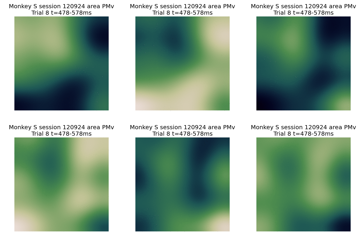
A video of the LFP waves that we analyzed:
The video shows the LFP (≤250 Hz) oscillation on all channels from a triple multi-electrode array implant, during one trial of the Cued Grasp with Instruct Delay task (see also Vargas-Irwin et al. 2015). Beta frequency (10-45 Hz) waves dominate, and occur in transients. The video is slowed down by a factor of 12.5. [get original original .avi video from Github]
Wave events in the phase plane
Some animated GIFs of β-LFP events:
Animated GIFs of some β-LFP events, showing filtered beta-LFP on each channel (left), enoised beta-LFP (middle), and extracted oscillation phase (right).


No comments:
Post a Comment